


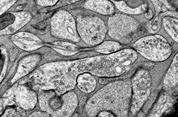
Flip open any neuroscience textbook and the depiction of a neuron will be roughly the same: a blobby, amoebalike cell body shooting out a long, thick strand. That strand is the axon, which conducts electrical signals to terminals where the cell communicates with other neurons. Axons have long been depicted as smooth and cylindrical, but a new study of mouse neurons challenges that view. Instead, it suggests their natural shape is more like a string of pearls. Even more provocatively, the authors propose those pearly bumps serve as control knobs, influencing how quickly and precisely the cell fires its signals.
The study, published today in Nature Neuroscience, should “100%” change how we’ve been thinking about neurons and their signals, says senior author Shigeki Watanabe, a molecular neuroscientist at John Hopkins University. Some outsiders agree. The findings are “highly significant and I think have been overlooked for quite some time,” says evolutionary biologist Pawel Burkhardt of the University of Bergen, who recently spotted similar pearl structures in neurons from tiny marine invertebrates known as comb jellies.
Yet several experts in the field contest the findings. Some cite potential confounding effects of the preparation and freezing method used to preserve cells before imaging. And some doubt the work totally upends what’s known about the true shape of the axon. “I think it’s true that [the axon is] not a perfect tube, but it’s not also just this kind of accordion that they show,” says neuroscientist Christophe Leterrier from Aix-Marseille University, who calls the study “a controversial addition to the literature.”
Since the mid-1960s, microscopists have seen that axons can scrunch up to form beads when they are diseased or under other stress. Leterrier has called these temporary beads “stress balls for the brain” and found evidence that they prevent cellular damage from spreading. Other studies suggest even normal axons bulge temporarily when cargo traveling to and from the cell nucleus forms a traffic jam, like the elephant bulging inside the body of a boa in the children’s book The Little Prince.
But Jacqueline Griswold, a graduate student in Watanabe’s lab, found something different in mouse neurons preserved with a technique called high-pressure freezing. The method, she says, can preserve “the fine structure of very, very small parts of cells” better than the chemical fixatives normally used in microscopy. She applied the technique both to mouse neurons grown in a dish and to samples from superthin slices of adult and embryonic mouse brain. (The team studied a subset of neurons that are unmyelinated, meaning they’re not wrapped up in an outer, insulating fatty layer that may constrain axon shape.)
Using an electron microscope, she noticed evenly spaced small pearls about 200 nanometers in diameter along the mouse axons. The blobs were smaller and more regularly spaced than Leterrier’s stress balls, and they didn’t contain anything, suggesting they didn’t result from cellular traffic jams.
The discovery echoes others: Watanabe had noticed similar pearled axons in roundworms in 2013 but didn’t investigate further, and Burkhardt recently saw them in comb jellies. Griswold also says she saw axon pearls in human brain tissue processed with high-pressure freezing, although she and her colleagues haven’t published that finding yet.
“The [beaded] shape itself is not surprising,” says Pramod Pullarkat, a biophysicist at the Raman Research Institute. He has studied the forces involved in axonal beading, and says a growing body of research suggests this shape can appear because of “pearling instability”—a physical phenomenon by which a cylindrical vesicle under tension scrunches up. It “may very well be” that this pearled shape is the normal state of axons, he says, but more research is needed to come to a definitive answer.
The pearling phenomenon is likely conserved over millions of years, Burkhardt says. He thinks the pearling probably has a functional purpose, and the new experiments offer a clue.
The speed at which an electrical signal travels along an axon depends on a complex interplay of its shape, diameter, and pearling pattern. Watanabe’s team modeled this interplay mathematically and also recorded the speed of signal conduction in real mouse neurons with different degrees of pearling. In both cases, they found that signals were slower when axon pearls were smaller and more tightly spaced, whereas wider spacing between pearls led to faster signals. This suggests the brain may change the subtle shape of neurons to improve signaling when it needs to compute a large amount of information, Watanabe says. But how exactly the brain might control pearling is not yet clear.
“Axon morphology in living animals is under constant and dynamic change,” says neuroscientist Tong Wang from ShanghaiTech University, who has studied beading in diseased neurons. She’s not surprised that axon beads could affect signal conduction and calls the finding a “compelling” discovery that adds to the mechanisms that make the brain so adaptable.
But other experts argue the pearls Watanabe’s team spotted are a side effect of cell damage. “While quick freezing is an extremely rapid process, something may happen during the manipulation of the sample … that also causes the beading,” says neuroscientist Pietro De Camilli from the Yale School of Medicine, who was not involved in the study. Cell biologist John Heuser from the Washington University School of Medicine in St. Louis also thinks beading is an artifact of the preparation process. “Axons can get to be that way … but they can also recover from it, they can go right back to looking like normal tubes.”
Griswold, however, says she also documented pearls in live neurons imaged with a technique that doesn’t require either freezing or chemical fixatives. And she showed that the paraformaldehyde used to prepare samples for standard microscopy introduces its own artifacts: Applying it causes the pearls to disappear. That could explain why previous studies documented a more cylindrical shape, she says.
“There have definitely been people who are like, ‘No, this cannot possibly be true,’” Griswold says. But, she adds, “We’ve published all of our experiments—check it out for yourself.”
Ultimately, these findings “open the door” for a better understanding of all different types of axon pearls, says biophysicist Joshua Zimmerberg from the National Institutes of Health. But to him, the big question is still open. “‘[Is pearling] affecting my thought processes?’—That’s what everybody would like to know.”
Without a way to observe the fine-grained structures of neurons while they’re active in a living human brain, “we don’t have powerful enough technology to answer it directly,” Zimmerberg says. For now, the work from Watanabe’s team is “as close as it gets.”
More: https://www.science.org/content/article/controversial-study-redraws-classical-picture-neuron
